
Segments of the ascending and descending aorta. rPA = right pulmonary
The aorta is divided into three separate zones for the purposes of REBOA (aortic length varies between individuals) Zone I of the aorta extends from the origin of the left subclavian artery to the coeliac artery (approx 20cm long in a young adult male) Zone II extends from the coeliac artery to the most caudal renal artery (approx 3cm long)

Aortic zones related to REBOA. Download Scientific Diagram
In a series of CT scans of the aorta from 100 patients diagnosed with severe aortic stenosis planned for transcatheter aortic valve implantation, we measured the intravascular distance from the.
Angiotomography with multiplanar and threedimensional reconstruction
Zone 2: From the celiac trunk to the lowest renal artery Zone 2 is an unused zone because if of difficulty in occluding the bleeding vessel at this aortic location; Zone 3: From lowest renal artery to the aortic bifurcation; Indications. Non-compressible hemorrhage below the diaphragm in the abdomen, pelvis or retroperitoneum

Schematic drawing outlining the three "landing zones" (green) for the
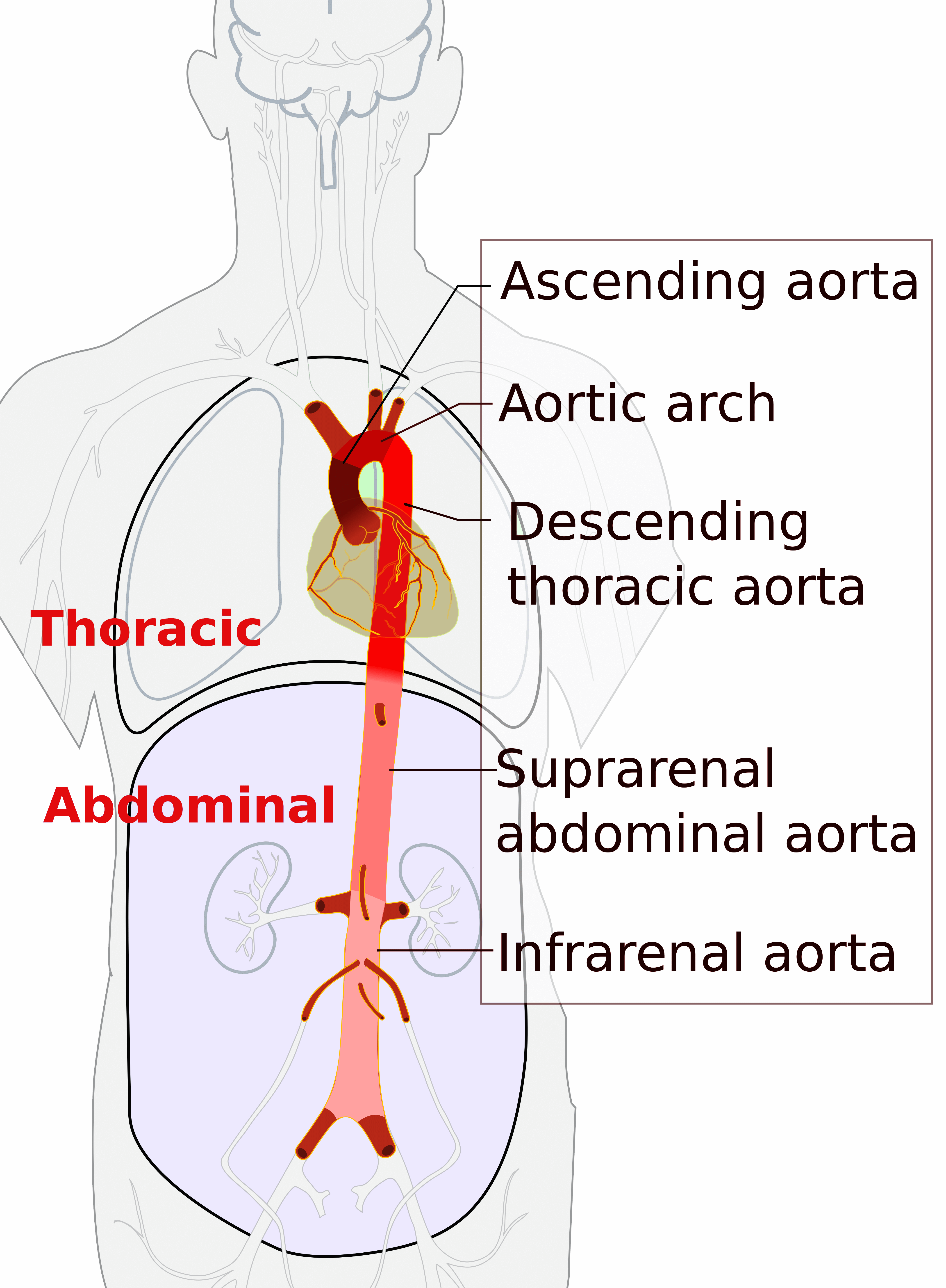
The aorta ( / eɪˈɔːrtə / ay-OR-tə; pl.: aortas or aortae) is the main and largest artery in the human body, originating from the left ventricle of the heart, branching upwards immediately after, and extending down to the abdomen, where it splits at the aortic bifurcation into two smaller arteries (the common iliac arteries ).

Thoracic Endovascular Aortic Repair Anesthesiology Clinics
The aorta is divided into three zones (Figure 1). Proper placement of the REBOA catheter is in either Zone I or Zone III. Zone I is located just distal to the take-off of the left subclavian artery, and ends at the celiac trunk. Zone III is located just distal to the lowest renal artery and ends at the aortic bifurcation. Zone II is

Proximal landing zones for aortic arch and upper descending thoracic
Of all injuries sustained in trauma, the aortic injury is one of the most time-sensitive, life-threatening conditions, second only to head injury as a cause of death. The morbidity and mortality associated with traumatic aortic injury are about 30% within the first 24 hours.

Aortic Zones A Surgeon's Notes
Small volume aortic occlusion balloons (AOB) have poor occlusion rates in zone I (0-2.8%) and III (4.4-34.4%). Conclusions Men and older age groups have longer CLL distances to zone I and III and introduction depths of AOB must be adjusted.

New Aortic Dissection Classification and Practical RealWorld
F. Zone 3 (first 2 cm. distal to left subclavian) G. Zone 4 (end of zone 3 to mid descending aorta ~ T6) H. Zone 5 (mid descending aorta to celiac) I. Zone 6 (celiac to superior mesenteric) J. Zone 7 (superior mesenteric to renals) K. Zone 8 (renal to infra-renal abdominal aorta) L. Zone 9 (infrarenal abdominal aorta) M. Zone 10 (common iliac)
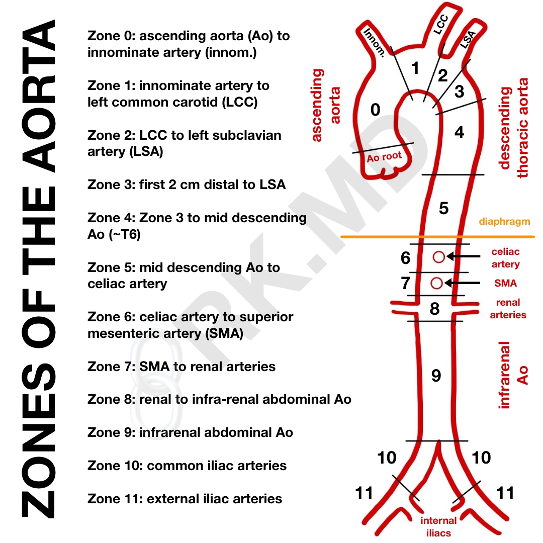
Zones Of The Aorta RK.MD
The aortic arch is a continuation of the ascending aorta and begins at the level of the second sternocostal joint. It arches superiorly, posteriorly and to the left before moving inferiorly. The aortic arch ends at the level of the T4 vertebra.

Society for Vascular Surgery (SVS) and Society of Thoracic Surgeons
Zone 2 continues from the celiac artery to the renal artery. Zone 3 extends from the origin of the lowest renal artery to the aortic bifurcation (infrarenal aorta). Zone 1 occlusion is utilized in patients in cardiac arrest or those in hemorrhagic shock with evidence of non-compressible hemorrhage arising below the diaphragm.

Aortic Anatomy and Complications of the Proximal Sealing Zone after
as aortic dissection and its variants (e.g., intramural hematoma [IMH]), rupture of ascending aortic aneurysm, aortic trauma, and penetrating ulcer.Otherentities,suchasTakayasuaortitis(TA),giant-cell(temporal) arteritis(GCA),andmycoticaneurysm,arediscussedbriefly.Lesscom-mon aortic diseases such as aortic tumors (because of their rarity) and

Ultrasound examination zones for the thoracic aorta. The ascending
Surgeons (SVS/STS) Aortic Dissection Classification System of dissection subtype according to zone location of pri-mary entry tear. Fig 4. An aortic dissection with an entry tear in zone 0 is classified as type A. In the example illustrated, the dissection process extends distally to zone 9, such that the dissection is fully classified as A 9.

Aortic Anatomy and Complications of the Proximal Sealing Zone after
Aortic dissection Type B Classification Aorta Dissection Reporting Section 1. Introduction Purpose of the document Acute aortic dissection is the most common emergency affecting the human aorta, with high mortality and morbidity without appropriate and time-sensitive treatment.
Society for Vascular Surgery/Society of Thoracic Surgeons (SVS/STS
When thoracic and thoracoabdominal aortic pathology pushes the limits of currently available thoracic endovascular aortic repair (TEVAR) technology, we need to enhance our techniques to repair increasingly complex anatomy that, if treated by open surgery, is associated with significant morbidity and mortality. 1 Compromised distal landing zones have been implicated in late type Ib endoleaks.
Aortic Dissection Explained Risk Factors, Symptoms, Diagnosis, FAQs
Zones Of The Aorta By Rishi November 22, 2021 0 As a cardiothoracic anesthesiologist and intensivist, I care for many patients in the OR and ICU who have aortic aneurysms or dissections undergoing open/endovascular repair.

Proximal landing zones for aortic arch and upper descending thoracic
Aortic regurgitation (AR) is the third most frequently encountered valve lesion and may be caused by abnormalities of the valve cusps or the aorta. Echocardiography is instrumental in the assessment of AR as it enables the delineation of valvular morphology, the mechanism of the lesion and the grading of severity. Severe AR has a major impact on the myocardium and carries a significant risk of.